


Under the Light Microscopic structure
In anatomy and histology, the identification of muscle tissues—skeletal, smooth, and cardiac—under a light microscope is crucial for understanding their structure and function. Each type of muscle tissue has distinct characteristics that can be identified through careful observation. Below is a detailed comparison and identification guide for these muscle tissue

1. Skeletal Muscle

Structure:
- Appearance: Skeletal muscle fibers are long, cylindrical, and multinucleated, with nuclei located at the periphery of the cells.
- Striations: These muscles exhibit a distinct striated (striped) appearance due to the organized arrangement of actin and myosin filaments.
- Arrangement: The fibers are arranged in parallel bundles, making them easy to identify.
Key Features of Skeletal Muscle Tissue:
Striations:
- The most prominent feature of skeletal muscle tissue is the presence of striations, visible as alternating light and dark bands across the muscle fibers. These striations are due to the organized arrangement of actin and myosin filaments within the muscle cells, which are responsible for muscle contraction.
Muscle Fibers:
- The muscle fibers in the slide are long, cylindrical, and run parallel to each other. These fibers are multinucleated, meaning each muscle cell has multiple nuclei.
Nuclei Placement:
- Although not clearly visible in this particular image, typically in skeletal muscle, the nuclei are located at the periphery (edges) of the muscle fibers, which is another distinguishing feature from other types of muscle tissue.
Fiber Bundles:
- The slide shows several bundles of muscle fibers, which is typical of how skeletal muscle tissue is organized. Each bundle is surrounded by connective tissue.
Slide Identification Points:
- Striations: Under the light microscope, look for alternating dark and light bands, which are the characteristic striations of skeletal muscle.
- Nuclei Position: The peripheral placement of nuclei is a key identifying feature.
- Fiber Size: Skeletal muscle fibers are generally larger and more uniform in size compared to other muscle types.
2. Smooth Muscle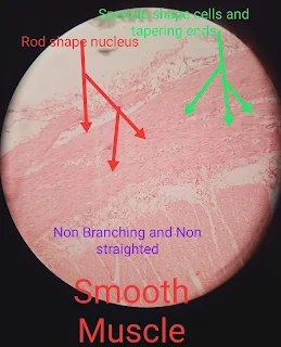

Structure:
- Appearance: Smooth muscle cells are spindle-shaped with a single, centrally located nucleus. Unlike skeletal muscle, smooth muscle lacks striations.
- Arrangement: The cells are arranged in sheets or layers, often found in the walls of hollow organs like the intestines and blood vessels.
Key Features of Smooth Muscle Tissue:
Spindle-Shaped Cells:
- The smooth muscle cells, also known as myocytes, are spindle-shaped with tapering ends. This shape allows them to fit together closely, forming a smooth, continuous sheet of muscle tissue. The image clearly labels and highlights these spindle-shaped cells.
Non-Striated Appearance:
- Unlike skeletal and cardiac muscle, smooth muscle lacks visible striations (bands). The slide shows a uniform appearance without the alternating light and dark bands seen in striated muscles, confirming it as smooth muscle tissue.
Rod-Shaped Nucleus:
- The nuclei of smooth muscle cells are centrally located and rod-shaped. In the image, the nuclei are elongated and aligned with the long axis of the muscle cells. The label correctly identifies these rod-shaped nuclei.
Non-Branching Fibers:
- Smooth muscle fibers do not branch, unlike cardiac muscle fibers. The image shows long, non-branching muscle fibers, which is typical of smooth muscle tissue.
Function and Location:
- Smooth muscle is primarily involved in involuntary movements, such as the contraction of blood vessels, the digestive tract, and other hollow organs. It is controlled by the autonomic nervous system.
Slide Identification Points:
- Lack of Striations: The absence of striations is a key feature distinguishing smooth muscle from skeletal and cardiac muscle.
- Nuclei: The centrally located, elongated nuclei are often visible under the light microscope.
- Cell Shape: The spindle-shaped, tapering ends of smooth muscle cells are also indicative.
- 3. Cardiac Muscle
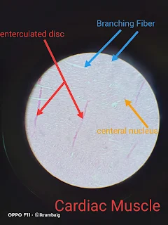

Structure:
- Appearance: Cardiac muscle cells are striated, like skeletal muscle, but they are shorter, branched, and interconnected. Each cell typically has one or two centrally located nuclei.
- Intercalated Discs: Unique to cardiac muscle are the intercalated discs, which appear as dark, irregular lines between the cells and are crucial for synchronizing heart contractions.
Key Features of Cardiac Muscle Tissue:
Branching Fibers:
- Cardiac muscle fibers are known for their branching structure. This branching allows for the formation of a complex network, which is crucial for the coordinated contraction of the heart. The image clearly labels and highlights these branching fibers.
Intercalated Discs:
- One of the hallmark features of cardiac muscle is the presence of intercalated discs. These structures are specialized connections between cardiac muscle cells that facilitate synchronized contraction. The intercalated discs appear as dark lines running across the muscle fibers and are clearly identified in the image.
Central Nucleus:
- Unlike skeletal muscle, where nuclei are located at the periphery of the fibers, cardiac muscle cells typically have a centrally located nucleus. The image shows these central nuclei, which are a distinguishing feature of cardiac muscle tissue.
Striated Appearance:
- Similar to skeletal muscle, cardiac muscle has a striated appearance due to the arrangement of actin and myosin filaments. Although the striations may not be as prominent as in skeletal muscle, they can still be observed under a light microscope.
Function and Location:
- Cardiac muscle is found exclusively in the heart and is responsible for pumping blood throughout the body. It is an involuntary muscle, meaning it contracts without conscious control, driven by the heart's own electrical system.
Slide Identification Points:
- Striations and Intercalated Discs: The combination of striations and the presence of intercalated discs is a defining feature of cardiac muscle.
- Cell Shape: Cardiac muscle cells are shorter and branched, which helps in distinguishing them from the long, cylindrical skeletal muscle fibers.
- Nuclei: The centrally located nuclei, often surrounded by a clear space, can help identify cardiac muscle under a microscope.
- Understanding the microscopic differences between skeletal, smooth, and cardiac muscle is essential in anatomy and histology. By observing features such as striations, cell shape, and nuclei placement, these tissues can be accurately identified under a light microscope.


0 Comments